Call Us Today!
(970) 247-2677
New Patients
(970) 233-7384


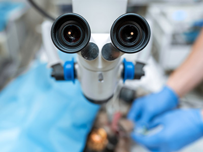
Dental care increasingly relies on technologies that reveal details the naked eye cannot. The 3D Seiler surgical microscope is a sophisticated visualization system that combines high-resolution optics with stereoscopic imaging to present depth and clarity during procedures. Rather than relying on two-dimensional images or limited magnification, Dr. Miner can operate with a true sense of scale and spatial relationships, which matters when working in the confined, delicate spaces inside a tooth.
Built for both precision and comfort, the system integrates adjustable magnification, superior illumination, and an intuitive eyepiece arrangement that preserves natural posture. Its 3D-capable camera records and projects what Dr. Miner sees, making documentation and case review more straightforward. Those capabilities help Dr. Miner make finer distinctions between healthy and compromised tissue while planning and executing targeted treatments.
At Mountain Health Dentistry, Dr. Miner uses advanced visualization tools like this microscope to support conservative, tooth-preserving care. When anatomy can be seen more clearly, it often allows for less tissue removal, more accurate targeting of infection, and treatment decisions that favor long-term function and stability.
Unlike standard dental loupes or basic microscopes, the 3D Seiler system delivers stereoscopic depth perception and a wide range of magnification without compromising field of view. Its optical train is engineered to minimize distortion and maximize contrast so that tiny structures—such as accessory canals, hairline fractures, or calcifications—are easier to identify. The end result is a clearer operational field that supports meticulous technique for Dr. Miner.
Not every visit to the dental office requires a surgical microscope, but there are many scenarios where its use significantly improves diagnostic accuracy and treatment quality. Endodontic procedures, including primary root canal therapy and retreatments, often demand the identification of additional canals, previously missed anatomy, or persistent infection pathways—situations in which Dr. Miner benefits greatly from magnification and depth perception.
Periodontal surgeries and microsurgery around the root tip (apical surgery) also benefit from the fine control the microscope affords Dr. Miner. For these procedures, seeing the precise margins of tissue, identifying the exact location of defects, and differentiating between inflamed and healthy tissue can change the course of treatment and support more conservative approaches.
The microscope is likewise helpful during restorative work where precise margins and fit are crucial—such as delicate crown preparations or complex repairs performed by Dr. Miner. By enabling more exact work, more natural tooth structure can often be preserved, reducing the chance of future complications.
Dr. Miner commonly uses the 3D Seiler system for detailed endodontic procedures, surgical interventions that require micro-dissection, and restorative cases where margin refinement is essential. It can assist in detecting fractures, locating calcified canals, and confirming the completeness of root canal obturation. In short, whenever depth perception and fine detail change the outcome, microscopy plays a role in Dr. Miner’s care.
A key advantage of the 3D microscope is improved diagnostic confidence for Dr. Miner. Some dental problems present with subtle visual cues—micro-fractures, tiny recurrent decay at restoration margins, or accessory canals—that are easy to miss without enhanced magnification. With clearer visualization, Dr. Miner can make more informed decisions about the necessity and extent of intervention.
The microscope also supports documentation and collaboration. High-quality 3D images and video allow Dr. Miner to review cases off-line, consult with colleagues, and track healing over time. For patients, visual evidence can make treatment explanations more transparent and help them understand recommendations more clearly.
In complex cases—such as retreatments where previous therapy failed—microscopic inspection by Dr. Miner frequently reveals the cause of persistent symptoms. By identifying exact problem areas, treatment can be more targeted, avoiding unnecessary removal of healthy tissue.
Enhanced illumination and magnification expose subtle anatomic features like isthmuses, accessory canals, and micro-cracks. These discoveries can alter treatment strategy in real time for Dr. Miner. For example, locating a previously undetected canal can mean the difference between a successful root canal and persistent infection.
Although the microscope is primarily a clinical tool, its use by Dr. Miner can translate into a better patient experience. Procedures guided by enhanced visualization tend to be more conservative, which can mean less postoperative discomfort and a reduced risk of complications. When work is completed precisely, patients often benefit from fewer follow-up interventions.
From the operator’s perspective, the ergonomic design of modern surgical microscopes reduces physical strain for Dr. Miner during detailed procedures, helping maintain consistent performance across longer appointments. Better ergonomics and clearer visuals support smoother, more predictable care.
That said, the primary goal remains clinical excellence rather than speed: Dr. Miner uses advanced visualization to ensure treatment is performed thoroughly and correctly, prioritizing long-term oral health and function.
Integrating a 3D surgical microscope into a dental practice requires attention to workflow, staff training, and sterile technique. The device becomes part of a broader system used by Dr. Miner and the team—paired with digital imaging, intraoral cameras, and restorative protocols—to deliver coordinated care.
The microscope’s recording capabilities also support interdisciplinary communication. Dr. Miner can archive high-resolution visual records for treatment planning or share files with specialists when collaborative care is needed. Shared visual information improves diagnostic consistency and clarity across cases.
At Mountain Health Dentistry, Dr. Miner prioritizes technologies that integrate smoothly into clinical workflow so that advanced visualization consistently translates into better patient outcomes.
Using the microscope effectively requires training, proper sterilization protocols, and attention to detail. Dr. Miner follows strict draping and disinfection procedures to maintain a sterile field while preserving image quality. Comprehensive record-keeping supports continuity of care and allows long-term evaluation of treatment success.
Recording cases also supports ongoing professional development, allowing Dr. Miner to refine techniques based on objective visual feedback. For patients, this documentation enhances transparency and supports informed decision-making.
If you’d like to learn more about how 3D surgical microscopy affects diagnosis, treatment planning, or the patient experience, please contact us. Dr. Miner and the team are happy to explain how advanced visualization supports conservative, precise dentistry focused on long-term oral health.