Call Us Today!
(970) 247-2677
New Patients
(970) 233-7384


Mountain Health Dentistry uses cone-beam computed tomography (CBCT) to bring three-dimensional clarity to dental diagnosis and treatment planning. CBCT captures detailed images of teeth, bone, nerves, and sinuses in a single, focused scan, giving clinicians a complete view of the structures that matter most when planning complex care. This technology helps translate clinical findings into safer, more predictable treatment paths without relying only on traditional two-dimensional X-rays.
By integrating CBCT into our diagnostic toolkit, we reduce guesswork and create more precise treatment plans tailored to each patient's anatomy. The images generated are clear enough to reveal subtle differences in bone volume, root anatomy, and spatial relationships—information that frequently changes how clinicians approach restorative, surgical, or endodontic care. For patients, that means fewer surprises and a higher likelihood of successful outcomes.
CBCT systems produce volumetric images that show anatomy from any angle. Instead of interpreting overlapping structures on flat films, clinicians can examine cross-sections, panoramic reconstructions, and 3D renderings to understand how teeth relate to surrounding bone and vital structures. This perspective is especially valuable when conventional X-rays provide incomplete or ambiguous information.
The level of detail captured in a CBCT scan helps identify anatomic variations—like extra canals, atypical root curvature, or bone defects—that might otherwise go unnoticed. Detecting these variations early enables the team to choose techniques and instruments that protect natural tooth structure and reduce the risk of complications during treatment.
Because images are digital, they can be measured and annotated with precision. Measurements of bone height, cortical thickness, and distances to nerves or sinuses become part of the clinical record and guide decisions such as implant size, surgical approach, or the need for tissue grafting. The result is a data-driven plan backed by visual evidence rather than assumptions.
One of the most transformative uses of CBCT is preoperative planning for implant placement and other oral surgeries. A 3D assessment reveals the true volume and quality of bone at potential implant sites and maps critical anatomy—like the inferior alveolar nerve and maxillary sinuses—so implants can be positioned safely and predictably.
Plan-driven workflows often use CBCT scans to create surgical guides or digital mockups, helping clinicians place implants at the ideal angle and depth for long-term stability and optimal prosthetic support. This coordinated approach minimizes intraoperative adjustments and helps ensure prosthetic components fit properly from the start.
For complicated extractions or apical surgeries, CBCT clarifies relationships between roots and adjacent structures, enabling more conservative and targeted interventions. When surgery is unavoidable, that preoperative insight shortens operative time, improves precision, and supports quicker recovery for patients.
CBCT is highly effective for investigating suspected pathology or unusual symptoms that aren’t explained by routine exams. It reveals lesions, cysts, and bony changes with greater sensitivity than standard radiographs, allowing clinicians to determine whether a problem is localized to an individual tooth or reflects a broader anatomic condition.
In addition to detecting pathology, CBCT aids evaluation of temporomandibular joint (TMJ) anatomy, impacted teeth, and the relationship of unerupted teeth to adjacent roots. Sinus anatomy is also visible, which is valuable when dental disease or implants interact with maxillary sinuses. Having these details helps clinicians coordinate care with oral surgeons, ENT specialists, or other providers when necessary.
Airway and sleep-related assessments can also benefit from three-dimensional imaging when clinically indicated. While CBCT is not a stand-alone diagnostic tool for sleep disorders, it provides objective information about airway volume and bony constraints that may be useful as part of a broader evaluation.
Modern CBCT units are designed to capture high-resolution images using focused fields of view (FOV), so the scan can be limited to the region of interest rather than exposing the entire head. This targeted approach adheres to the principle of keeping radiation “as low as reasonably achievable” while still providing diagnostic-quality images.
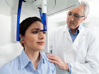
Scans typically take only seconds to acquire, and the open design of most CBCT machines is less confining than some medical imaging devices. Patients remain seated or standing in a stable, supported position, which helps reduce motion artifact and improves image clarity. For many people, the experience is quick and well tolerated.
In addition to limiting exposure, clinicians use CBCT selectively: images are ordered only when the expected diagnostic benefit outweighs the minimal risk. When a 3D scan meaningfully changes the treatment approach or increases procedural safety, the information it provides often justifies its use.
CBCT data integrates smoothly with other digital tools—like intraoral scans, CAD/CAM systems, and surgical guide software—creating cohesive digital treatment plans. This interoperability improves communication between the dental team and laboratory partners, and it streamlines the process from diagnosis to final restoration.
When cases require specialty input, CBCT volumes can be shared securely with oral surgeons, periodontists, or radiologists for collaborative planning. That shared view of the anatomy reduces miscommunication and supports coordinated care, which benefits patients who need multidisciplinary treatment.
For patients, integration means clearer explanations of recommended procedures. Visualizing the 3D anatomy on screen helps clinicians show where treatment is needed and why certain options are preferred, empowering patients to make informed decisions about their care.
CBCT is a powerful diagnostic tool that supports safer, more predictable dental care when used thoughtfully and selectively. Mountain Health Dentistry applies this technology to enhance clinical judgment, improve surgical planning, and communicate treatment options clearly—always with an emphasis on patient comfort and evidence-based decision making. If you’d like to learn more about how CBCT might be used in your care, please contact us for more information.