Call Us Today!
(970) 247-2677
New Patients
(970) 233-7384


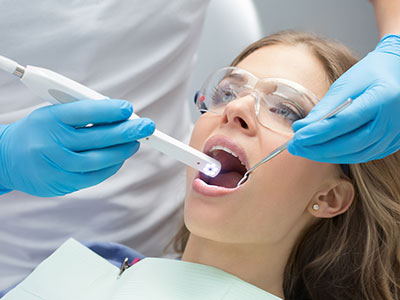
An IOS (intraoral scanner) is a handheld digital device that captures a detailed three-dimensional record of the teeth and surrounding oral tissues. Instead of producing a physical impression with trays and putty, Dr. Miner uses the scanner to record a continuous stream of images that are stitched together in real time to create a highly accurate virtual model. These models show surface texture, margins, and soft-tissue contours with fine resolution, making them useful for diagnostics, restorative design, and long-term records.
Modern scanners combine optical sensors, LED illumination, and advanced software algorithms to translate optical data into precise digital files. The resulting 3D captures are immediately viewable on a monitor, allowing Dr. Miner to inspect occlusion, identify undercuts, and verify margins from multiple angles. Because the data is digital from the start, it can be trimmed, annotated, and exported in standard formats such as STL or PLY for further processing by dental labs and design software.
Beyond impressions, IOS devices collect information that supports comprehensive clinical decision-making for Dr. Miner. Color mapping and overlays can highlight wear facets or areas of demineralization, while integrated measurement tools provide exact distances and angles. The real-time feedback loop between the scanner and Dr. Miner reduces guesswork and elevates the level of precision available during same-day restorations or complex treatment planning.
Digital impressions from an IOS scanner become the foundation for a more informed treatment plan. Because scans are accurate and reproducible, they enable closer coordination between Dr. Miner, dental laboratories, and specialists. For example, a high-resolution file can be sent to a lab for CAD/CAM milling or shared with a prosthodontist or orthodontist using objective digital data rather than relying on physical casts that may warp or deteriorate over time.
IOS data integrates smoothly with other digital tools—CBCT scans, intraoral photos, and practice management systems—allowing Dr. Miner to layer information and see relationships that aid diagnosis. This interoperability supports better case acceptance and helps simulate outcomes before irreversible steps are taken. Digital planning tools also allow Dr. Miner to preview restorations, check occlusal contacts virtually, and adjust designs to conserve tooth structure while achieving predictable results.
Because scans are stored electronically, Dr. Miner can compare records over time to monitor changes in tooth position, wear, or gingival recession. This longitudinal view supports early intervention when patterns emerge and allows objective tracking of treatment outcomes. In short, intraoral scanning provides a precise, shareable, and durable dataset that improves the consistency and predictability of care.
From the patient’s perspective, intraoral scanning removes many of the unpleasant elements associated with traditional impressions. There is no need for bulky trays or impression materials that can cause gagging or discomfort; instead, Dr. Miner gently moves the scanner through the mouth while the patient watches a real-time reconstruction of their teeth on a screen. This visual feedback helps patients understand their condition and proposed treatment more clearly.
Speed is another benefit. Scans are often completed more quickly than conventional impressions, particularly for multi-unit restorations or full-arch captures. Because the data is immediately available, Dr. Miner can begin design workflows without delay, shortening the overall treatment timeline. For patients with sensitive gag reflexes or limited opening, the noninvasive nature of scanning can make visits more comfortable and less stressful.
The digital workflow also supports more patient-centered care. Dr. Miner can annotate scans chairside to explain findings, show how a restoration will fit, and involve patients in decision-making. This transparency builds trust and helps patients feel more informed about their options.
Precision matters when the goal is to preserve as much natural tooth structure as possible. High-resolution intraoral scans enable conservative preparations and restorations with tight marginal fit, which lowers the risk of recurrent decay or unnecessary reshaping. When restorations are designed from accurate digital impressions, Dr. Miner can achieve better fit on the first try, reducing repeated adjustments that may compromise tooth structure over time.
In restorative workflows—crowns, onlays, veneers, and implant restorations—the scanner supports predictable results by allowing Dr. Miner to evaluate interproximal contacts, occlusal relationships, and margin placement before fabrication. This is especially valuable in tooth-preserving procedures where small differences in fit can affect longevity. Our use of advanced scanning technology reflects a commitment to conservative care and long-term oral health.
Beyond individual restorations, IOS scans can be used by Dr. Miner to monitor periodontal changes and evaluate tissue response around crowns and implants. Documented digital records support maintenance planning and early intervention, helping extend the life of natural teeth and restorations alike.
Introducing intraoral scanning requires a structured clinical protocol to maximize accuracy and efficiency. Proper scanning technique, calibration, and ongoing team training ensure consistent results for Dr. Miner across patients and procedures.
Digital records are handled within encrypted systems and integrated with practice software according to patient data protection standards. This ensures that scans used by Dr. Miner are securely stored and only accessible to authorized members of the care team or external partners with consent.
Over time, this digital workflow supports measurable quality improvement. Because every restoration begins with a recorded scan, Dr. Miner can review outcomes, track adjustments, and refine protocols based on objective data. This continuous feedback loop strengthens clinical performance and enhances patient care.
In summary, intraoral scanning provides a precise, patient-friendly, and efficient approach to modern dentistry. By converting optical data into accurate digital models, IOS technology enhances diagnosis, treatment planning, and restorative outcomes while improving the patient experience. If you’d like more information about how intraoral scanning is used in our practice or how it may benefit your care, please contact us for further details.